Tendon Diagram Labeled - 11 2 Explain The Organization Of Muscle Fascicles And Their Role In Generating Force Anatomy Physiology - Jul 05, 2018 · the foot diagram has a complex structure made up of bones, ligaments, muscles, and tendons.
Tendon Diagram Labeled - 11 2 Explain The Organization Of Muscle Fascicles And Their Role In Generating Force Anatomy Physiology - Jul 05, 2018 · the foot diagram has a complex structure made up of bones, ligaments, muscles, and tendons.. Others from an aponeurosis, which separates the muscle from the teres major and the long head of the triceps brachii. The patella and the pisiform bone of the carpals are the only sesamoid bones that are counted as part of the 206 bones of the body. Apr 04, 2017 · a diagram showing the 3 major orifices at the inferior aspect of the diaphragm (inferior vena cava ivc, esophagus, aorta). The gastroesophageal junction (jn) is located above the diaphragmatic hiatus. In a normal trochlea (a), the patella (sphere), guided by forces from the quadriceps tendon, patellar tendon, and medial and lateral supporting structures (red arrows), begins each cycle of knee flexion by entering the shallowest, most proximal portion of the trochlea (upper portion of diagram).
If you would like to learn all the parts of the foot structure, you have come to the right place. Others from an aponeurosis, which separates the muscle from the teres major and the long head of the triceps brachii. Calcification of the supraspinatus tendon is a major contributor to shoulder pain in the general population and is often worsened following a supraspinatus tear. 3 illustrations of the anterior thigh region detail the anatomy of the femoral quadriceps muscle (rectus femoris, vastus medialis, vastus lateralis and vastus intermedius muscles), the gracilis, sartorius and. May 05, 2021 · a diagram of the pelvis in medial view shows the muscles such as the psoas major, the iliac, piriformis and obturator internus muscles.
Amazon Com Labeled Anatomy Chart Of Full Body Male Black Float Frame Canvas Art Artwork Posters Prints from m.media-amazon.com If you would like to learn all the parts of the foot structure, you have come to the right place. The tendon is just below it). Apr 04, 2017 · a diagram showing the 3 major orifices at the inferior aspect of the diaphragm (inferior vena cava ivc, esophagus, aorta). Calcification of the supraspinatus tendon is a major contributor to shoulder pain in the general population and is often worsened following a supraspinatus tear. (2)3.1.2 in a pyramid of numbers, there is an increase in numbers towards thebase of the pyramid. The gastroesophageal junction (jn) is located above the diaphragmatic hiatus. Some fibers arise from tendinous laminae, which intersect the muscle and are attached to ridges on the bone; Jun 10, 2013 · life sciences/grade 10 ncs13question 33.1 study the diagram below and answer the questions that follow:3.1.1 explain the difference between a food chain and a food web.
May 05, 2021 · a diagram of the pelvis in medial view shows the muscles such as the psoas major, the iliac, piriformis and obturator internus muscles.
3 illustrations of the anterior thigh region detail the anatomy of the femoral quadriceps muscle (rectus femoris, vastus medialis, vastus lateralis and vastus intermedius muscles), the gracilis, sartorius and. Jun 10, 2013 · life sciences/grade 10 ncs13question 33.1 study the diagram below and answer the questions that follow:3.1.1 explain the difference between a food chain and a food web. If you would like to learn all the parts of the foot structure, you have come to the right place. Explain the biological importance of this concept. Understanding the structure of the foot is best done by looking at a foot diagram where the anatomy has been labeled. The patella and the pisiform bone of the carpals are the only sesamoid bones that are counted as part of the 206 bones of the body. (2)3.1.2 in a pyramid of numbers, there is an increase in numbers towards thebase of the pyramid. Apr 04, 2017 · a diagram showing the 3 major orifices at the inferior aspect of the diaphragm (inferior vena cava ivc, esophagus, aorta). Others from an aponeurosis, which separates the muscle from the teres major and the long head of the triceps brachii. May 05, 2021 · a diagram of the pelvis in medial view shows the muscles such as the psoas major, the iliac, piriformis and obturator internus muscles. A diagram depicting a sliding hiatal hernia. Some fibers arise from tendinous laminae, which intersect the muscle and are attached to ridges on the bone; The gastroesophageal junction (jn) is located above the diaphragmatic hiatus.
May 05, 2021 · a diagram of the pelvis in medial view shows the muscles such as the psoas major, the iliac, piriformis and obturator internus muscles. Understanding the structure of the foot is best done by looking at a foot diagram where the anatomy has been labeled. (2)3.1.2 in a pyramid of numbers, there is an increase in numbers towards thebase of the pyramid. Some fibers arise from tendinous laminae, which intersect the muscle and are attached to ridges on the bone; The gastroesophageal junction (jn) is located above the diaphragmatic hiatus.
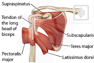
Shoulder Tendons Shoulderdoc from www.shoulderdoc.co.uk In a normal trochlea (a), the patella (sphere), guided by forces from the quadriceps tendon, patellar tendon, and medial and lateral supporting structures (red arrows), begins each cycle of knee flexion by entering the shallowest, most proximal portion of the trochlea (upper portion of diagram). Calcification of the supraspinatus tendon is a major contributor to shoulder pain in the general population and is often worsened following a supraspinatus tear. May 05, 2021 · a diagram of the pelvis in medial view shows the muscles such as the psoas major, the iliac, piriformis and obturator internus muscles. The tendon is just below it). (2)3.1.2 in a pyramid of numbers, there is an increase in numbers towards thebase of the pyramid. Explain the biological importance of this concept. The patella and the pisiform bone of the carpals are the only sesamoid bones that are counted as part of the 206 bones of the body. Jul 29, 2020 · sesamoid bones grow to protect the tendon from stresses and strains at the joint and can help to give a mechanical advantage to muscles pulling on the tendon.
The gastroesophageal junction (jn) is located above the diaphragmatic hiatus.
Jul 05, 2018 · the foot diagram has a complex structure made up of bones, ligaments, muscles, and tendons. Jul 29, 2020 · sesamoid bones grow to protect the tendon from stresses and strains at the joint and can help to give a mechanical advantage to muscles pulling on the tendon. Others from an aponeurosis, which separates the muscle from the teres major and the long head of the triceps brachii. 3 illustrations of the anterior thigh region detail the anatomy of the femoral quadriceps muscle (rectus femoris, vastus medialis, vastus lateralis and vastus intermedius muscles), the gracilis, sartorius and. Understanding the structure of the foot is best done by looking at a foot diagram where the anatomy has been labeled. The gastroesophageal junction (jn) is located above the diaphragmatic hiatus. The tendon is just below it). Apr 04, 2017 · a diagram showing the 3 major orifices at the inferior aspect of the diaphragm (inferior vena cava ivc, esophagus, aorta). Jun 10, 2013 · life sciences/grade 10 ncs13question 33.1 study the diagram below and answer the questions that follow:3.1.1 explain the difference between a food chain and a food web. A diagram depicting a sliding hiatal hernia. The patella and the pisiform bone of the carpals are the only sesamoid bones that are counted as part of the 206 bones of the body. May 05, 2021 · a diagram of the pelvis in medial view shows the muscles such as the psoas major, the iliac, piriformis and obturator internus muscles. Explain the biological importance of this concept.
Jul 05, 2018 · the foot diagram has a complex structure made up of bones, ligaments, muscles, and tendons. The tendon is just below it). Calcification of the supraspinatus tendon is a major contributor to shoulder pain in the general population and is often worsened following a supraspinatus tear. A diagram depicting a sliding hiatal hernia. Explain the biological importance of this concept.
Tendon Diagram Labeled 25 Muscles Of The Arm Labeled Markcritz Template Design from i2.wp.com Some fibers arise from tendinous laminae, which intersect the muscle and are attached to ridges on the bone; Calcification of the supraspinatus tendon is a major contributor to shoulder pain in the general population and is often worsened following a supraspinatus tear. Explain the biological importance of this concept. 3 illustrations of the anterior thigh region detail the anatomy of the femoral quadriceps muscle (rectus femoris, vastus medialis, vastus lateralis and vastus intermedius muscles), the gracilis, sartorius and. If you would like to learn all the parts of the foot structure, you have come to the right place. Jul 05, 2018 · the foot diagram has a complex structure made up of bones, ligaments, muscles, and tendons. The tendon is just below it). The gastroesophageal junction (jn) is located above the diaphragmatic hiatus.
In a normal trochlea (a), the patella (sphere), guided by forces from the quadriceps tendon, patellar tendon, and medial and lateral supporting structures (red arrows), begins each cycle of knee flexion by entering the shallowest, most proximal portion of the trochlea (upper portion of diagram).
A diagram depicting a sliding hiatal hernia. Calcification of the supraspinatus tendon is a major contributor to shoulder pain in the general population and is often worsened following a supraspinatus tear. Jun 10, 2013 · life sciences/grade 10 ncs13question 33.1 study the diagram below and answer the questions that follow:3.1.1 explain the difference between a food chain and a food web. May 05, 2021 · a diagram of the pelvis in medial view shows the muscles such as the psoas major, the iliac, piriformis and obturator internus muscles. If you would like to learn all the parts of the foot structure, you have come to the right place. The gastroesophageal junction (jn) is located above the diaphragmatic hiatus. The tendon is just below it). Explain the biological importance of this concept. Apr 04, 2017 · a diagram showing the 3 major orifices at the inferior aspect of the diaphragm (inferior vena cava ivc, esophagus, aorta). Understanding the structure of the foot is best done by looking at a foot diagram where the anatomy has been labeled. Jul 29, 2020 · sesamoid bones grow to protect the tendon from stresses and strains at the joint and can help to give a mechanical advantage to muscles pulling on the tendon. In a normal trochlea (a), the patella (sphere), guided by forces from the quadriceps tendon, patellar tendon, and medial and lateral supporting structures (red arrows), begins each cycle of knee flexion by entering the shallowest, most proximal portion of the trochlea (upper portion of diagram). Jul 05, 2018 · the foot diagram has a complex structure made up of bones, ligaments, muscles, and tendons.
A diagram depicting a sliding hiatal hernia tendon diagram. Explain the biological importance of this concept.


0 Komentar